What do we know about the mammogram used to detect breast cancer?
A Decrease font size. A Reset font size. A Increase font size.
The Bulletin of the Family Health Care Institute, King Hussein Foundation, today, Sunday, talks about mammograms, which are used for early detection of breast cancer in women who do not have any symptoms.
The institute's bulletin explains what a mammogram is, its types, and its common uses, in addition to how to prepare for this type of imaging, and recommended procedures before imaging.
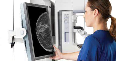
What is a mammogram? A mammogram is an X-ray examination of the breast, which is used to detect and diagnose breast diseases in women who have breast problems, such as swelling, pain or discharge from the nipple, as well as in women who do not have breast problems for the purpose of screening breast cancer. The procedure allows breast cancers, polyps, and cysts to be detected before they are detected by palpation. A normal mammogram cannot prove the presence of a cancerous mass, but if cancer is highly suspected, a tissue sample may be taken. Tissue can be taken with a special needle or an open surgical biopsy and examined under a microscope to determine if it is cancerous. Mammography has been used for about 30 years, and in the past 15 years, technical advances have greatly improved both technique and results. Today, dedicated equipment, used only for mammograms, produces high-quality studies and, at the same time, is low in radiation dose and therefore the radiation risks are minimal.
Types of mammograms: There are three recent developments in mammography, including the following types: 1. Digital mammography: Also called full field digital mammography, it is a system in which radiography examines the breast using low-dose X-rays with a specially designed breast implant that converts the X-rays into electrical signals. Electrical signals are used to produce images of the breast that can be seen on a computer screen, or printed on special film to look like a mammogram. These systems are similar to those in digital cameras and their efficiency allows for better images with a lower radiation dose. These images of the breast are transferred to a computer for review by the radiologist and for long-term storage. The patient experience during a digital mammogram is similar to that of a traditional mammogram.
2. Computer aided detection: Computer aided detection (CAD) systems look for areas of abnormal density, mass, or calcification on mammograms that may indicate the presence of cancer cells. This system highlights these areas in the images, and prompts the radiologist to carefully evaluate this area.

3. Breast tomosynthesis: A breast tomography, also called a three-dimensional (3D) mammogram, is an advanced form of breast imaging in which multiple images of the breast are taken from different angles and reconstructed (synthesized) in three a set of dimensional images. In this way, 3D mammography is similar to computed tomography (CT) Although the radiation dose for some breast CT systems is slightly higher than the dose used for standard mammography, it is still within FDA-approved safe levels of radiation from breast imaging Mammogram.
Large population-based studies have shown that CT screening improves breast cancer detection rates and reduces the number of cases in which women are recalled from screening for additional testing because of a possible abnormal finding.
A CT mammogram may also help with:- Early detection of small breast cancers that may be hidden in conventional mammography.- Fewer unnecessary biopsies or additional tests.- Greater likelihood of detecting multiple breast tumors.- Clearer images of abnormalities within high-density breast tissue. Greater accuracy in determining the size, shape, and location of breast abnormalities.
What are some common uses for this procedure? Mammograms are used as a screening tool for early detection of breast cancer in women who do not have any symptoms. It can also be used to detect and diagnose breast diseases in women who have symptoms such as swelling, tender skin, or nipple discharge.
1. Mammography screening for breast lumps: Mammography plays a key role in the early detection of breast cancers because it can show changes in the breast years before a patient or doctor notices them. Current guidelines from the American College of Radiology and the National Comprehensive Cancer Network (NCCN) recommend screening mammography every year for women, starting at age 40. Research has shown that annual mammograms result in the earliest detection of breast cancer that provides the most beneficial and maintenance treatment the breast.
The American College of Radiology (ACR) and the National Cancer Institute (NCI) also suggest that women with breast cancer, and those at increased risk due to a family history of breast or ovarian cancer, should seek professional medical advice about whether they should start In screening before the age of 40 or needing other types of screening. If you're at high risk of breast cancer, you may need to have a breast MRI in addition to your annual mammogram.
2. Diagnostic mammography: Diagnostic mammography is used to evaluate a patient with abnormal clinical findings such as a breast mass or nipple discharge.
How should I prepare for a mammogram? Before scheduling a mammogram, the American Cancer Society (ACS) and others recommend discussing any new breast findings or changes with your doctor. In addition, inform your doctor about any previous surgeries, hormone use, and family or personal history of breast cancer. Don't schedule your mammogram for the week before your period as hormonal changes affect the breasts during this time. The best time for a mammogram is a week after your period. Always tell your doctor or x-ray technician if there is any possibility that you may be pregnant.
The ACS also recommends the following before imaging: - Do not apply deodorant, powder or lotion under your arms or on your breasts on the day of the examination. These can show up on a mammogram as calcium spots. Describe any symptoms or breast problems to the technician performing the examination. Obtain your previous mammograms and make them available to the radiologist if they were done in a different location. This is required to compare with your current test and can often be obtained on CD. Ask when your results will be available. Don't assume results are normal if you haven't heard from your doctor.


