A new shooting technique that shows an unprecedented way ... video
A team of researchers developed and tested a new method of photography that they say will accelerate the research based on the laboratory by allowing researchers to take pictures of the blood vessels with different spatial standards..
The blood vessels are very important when it comes to the body's health function, and health researchers and professionals need to know the greatest possible place about the place where these small transport channels are heading..
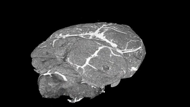
The newly developed three -dimensional visualization technique is called Vascuviz, which uses a fast -prepared polymer mix that fills the blood vessels and makes it visible to a variety of scanning techniques while moving around tissues and organs, according to "Russia Today".
The VASCUVIZ test was signed in mice tissue, which enabled researchers to photograph the vascular structure in the tissues, which could clarify the complex role of blood flow in health and disease, along with detailed sports models or complementary images of other tissue elements.
"The laboratory mice tests so far indicate that they work with different standards, from large arteries to the smallest capillaries, and can be used to show the details that traditional technologies may miss, which improves our understanding of howWork tissue ".
The measurements taken by VASCUVIZ can then enter into computer flow simulation operations, including cancer models, to learn how blood flow, which is necessary to understand how diseases work and the possibility of providing them.
What makes the new technology very useful is that it provides the approach of everyone in one, which provides the results and a level of details that usually take several scanning and several different ways to achieve.

Current photography methods, such as MRI, computerized tomography (CT) and microscopy, provide an important role in studying blood vessels in the laboratory, but it does not work together well and must be operated separately.
"Usually, if you want to collect data on the blood vessels into a specific tissue and integrate it with all the context surrounding it such as the structure and the types of cells that grow there, then you should renameFabric several times, get multiple pictures and collect supplementary information together ".
He continued: "This can be an expensive process and take a long time and threaten to destroy the tissue structure, which prevents us from using the collected information in new ways.".
Vascuviz technology uses a group of imaging factors: BRITEVU (used in computerized tomography), and Galbumin-Rhodamine (used in MRI tests).What is produced after that is a wonderful three -dimensional model.
While the researchers tested the technique of Vascuviz on mice only so far, they should work successfully on humans.In addition, the elements needed for them are available at reasonable prices.
Cancer tumors, leg muscles, brain, kidney tissue and periodic system can be photographed by Vascuviz, says the project responsible for the project.
The researchers pointed out that the combined vascular images should not only enhance the study of diseases that involve abnormalities in blood flow, such as cancer and stroke, but also enhance our understanding of tissue structures and functions throughout the body.
The team continued: "We hope that these developments will open in vascular photography before clinical, along with the new perception curricula presented here, new horizons for the biology of vascular image -based systems, and help to answer important questions in the broader field of the fine blood circulation and their role in health and disease".
Follow the health statement via Google News
طباعةEmailفيسبوك تويتر لينكدين Pin InterestWhats App


